Houston, Texas
Diagnostic Services
State-of-the-Art Technology
Millennium Diagnostic Center offers the latest technology, a highly-trained staff, and a relaxing, comfortable atmosphere in our medical imaging facilities. We partner with the leading imaging companies to bring the latest technology to our patients, ensuring the most accurate assessment and treatment. Our services allow physicians with resources to determine the effectiveness of the cancer treatment as well as the ability to monitor the recurrence.
Our diagnostic facilities are accredited by the American College of Radiology to perform a wide range of diagnostic imaging services. All examinations are performed by registered technologists and board-certified radiologists and interpreted by physicians who are certified by the American Board of Radiology. We have 8 convenient locations throughout the Greater Houston area – Kingwood, North Houston, Tomball, and The Woodlands. With an expert team by your side, we work together to determine the best treatment for you.
We are proud to offer a full range of advanced diagnostic imaging services:
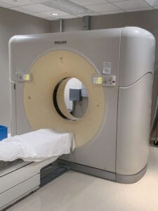
Diagnostic CT / Low-Dose Computer Tomography (CT)
A CT scan can provide multiple images inside the body using x-ray equipment or contrast material. Your doctor may recommend a CT scan to detect cancer or heart disease, internal injuries or for diagnosing muscle or bone disorders.
Preparation for your CT will depend on the type of exam. Before your CT scan, please contact us so we can provide specific instructions and review your health and insurance information.
Specialized CT Imaging Procedures Offered
- CT Angiographic Scans
- This type of test is an X-ray exam used to visualize the inside of blood vessels and organs within the body to look for signs of blockage or narrowing of the blood vessels that may be interfering with normal blood flow in the body.
- CT High Resolution Chest Scans
- CT Low Dose Lung Scans
- Scans are now available to high risk lung cancer patients. Click here for more information about lung cancer testing.
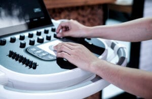
An echocardiogram is a non-invasive diagnostic test used to create detailed images of the heart, including its chambers, valves, walls, and the blood vessels that are connected to it, such as the aorta, arteries, and veins. It utilizes sound waves (ultrasound) to create images of the heart in real time, providing valuable information about the heart's size, shape, and function. Echocardiography is a safe and widely used diagnostic tool that provides valuable information about the heart and helps guide treatment decisions for a variety of heart conditions.
Echocardiograms are used for various purposes:
- Diagnosing Heart Conditions: They can help diagnose a wide range of heart conditions, including coronary artery disease, heart valve disease, heart failure, and congenital heart defects.
- Assessing Heart Function: Echocardiograms provide information about the heart's pumping capacity, the thickness and movement of its walls, and the condition of its valves.
- Monitoring Treatments: They are used to monitor the effectiveness of certain treatments, such as medication or surgery, and to track the progression of heart diseases over time.
- Guiding Interventions: In some cases, echocardiography is used during procedures such as heart catheterizations or valve repairs to guide the physician's actions.
Millennium Physicians offers a variety of echocardiograms to meet patients' needs, including the following types:
1.Transthoracic echocardiogram (TTE): This is the most common type of echocardiogram. A transducer (a small, handheld device) is placed on the chest to capture images of the heart from outside the body.
2.Transesophageal echocardiogram (TEE): A probe with an ultrasound transducer is passed through the mouth into the esophagus. This allows for a closer view of the heart, particularly the valves and structures behind the heart, as there are no lung or ribcage barriers.
3.Doppler echocardiogram: This type of echocardiogram uses Doppler ultrasound to measure the speed and direction of blood flow through the heart and blood vessels.
4.Stress echocardiogram: This test is performed before and after exercise (usually on a treadmill or stationary bike) to evaluate how the heart functions during increased activity.
Flow cytometry is a laboratory technique used to analyze and sort cells based on their physical and chemical characteristics. It uses a laser to illuminate and measure the properties of individual cells or particles as they pass through a narrow tube, allowing for the rapid analysis of a large number of cells. This technology is widely used in a range of research and clinical applications, including:
1.Immunophenotyping: Flow cytometry can be used to characterize the immune cells present in a blood sample. By staining cells with fluorescently labeled antibodies that bind to specific cell surface markers, researchers can identify and quantify different subsets of immune cells, such as T cells, B cells, and natural killer cells.
2.Cell Cycle Analysis: Flow cytometry can also be used to study the cell cycle, the process by which cells grow, replicate their DNA, and divide. By staining cells with fluorescent dyes that bind to DNA, researchers can determine the percentage of cells in each phase of the cell cycle (e.g., G0/G1, S, G2/M), providing insight into the proliferation and growth characteristics of a cell population.
3.Apoptosis Detection: Flow cytometry can be used to assess cell viability and detect programmed cell death (apoptosis). Cells that are undergoing apoptosis often exhibit changes in their cell surface markers or membrane permeability, which can be detected using fluorescent probes. This allows researchers to quantify the percentage of apoptotic cells in a population and study the mechanisms of apoptosis in different cell types.
4.Cancer Diagnostics: Flow cytometry is commonly used in the diagnosis and classification of hematological malignancies (blood cancers) such as leukemia and lymphoma. By analyzing the expression of specific cell surface markers, flow cytometry can help identify the type of cancer and determine the prognosis of the disease.
5.Stem Cell Analysis: Flow cytometry can be used to isolate and characterize stem cells, which are undifferentiated cells with the potential to differentiate into different cell types. By staining cells with fluorescently labeled antibodies that target specific stem cell markers, researchers can identify and purify stem cell populations for further study or therapeutic applications.
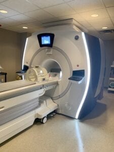
MRI (Magnetic Resonance Imaging)
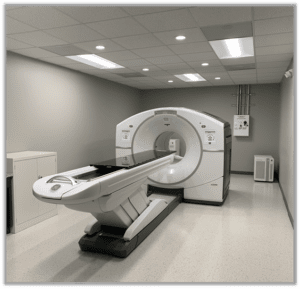
An MRI is an imaging test that uses radio waves and powerful magnetic fields to produce detailed, computer-generated pictures of areas inside the body. These pictures can show the difference between healthy and unhealthy tissues. Unlike X-rays, an MRI does not use radiation and deliveries better images for in-depth analysis.
The MRI scanner at Millennium Diagnostic Center provides safe, accurate diagnostic imaging for a wide array of injuries, conditions and diseases. An MRI has multiple diagnostic purposes:
- Determine if a tumor is noncancerous or cancerous
- Learn more about the size and location of the tumor
- Help plan cancer treatments, such as surgery or radiation therapy
- Monitor how well treatment is working
- Diagnose cardiac conditions and strokes
- Assess blood flow and vascular conditions
- Diagnose breast disease
- Examine male and female reproductive organs
- Determine and examine conditions and injuries to the brain, neck, spinal cord, bones, joints and other soft tissues
- Evaluate infections
Our MRIs are designed to help maximize patient comfort while maintain excellent image quality. The new GEM Suite surface coil technology reflects the importance of making the geometry of the equipment and technology compatible with the patient. The combined features of the entire Suite are designed to facilitate high resolution, signal-to-noise (SNR) imaging from the top of the head down to the feet, while maximizing the comfort of patients across many different shapes, sizes and situations.
The MRI is performed by a radiology technologist who is specially trained and certified to perform an MRI. After the MRI is completed, our highly experienced, board-certified radiologists read and interpret the imaging tests. A signed report will be sent to your primary care or referring physicians, who will share results with you.
Millennium Physicians is proud to offer an advanced new MR solution for the analysis, planning, and targeted biopsy of the prostate. Magnetic Resonance/Ultrasound (MR/US) fusion biopsy is a technique that combines MRI imaging with real-time ultrasound to guide the sampling of prostate tissue for analysis.
MR/US fusion biopsy has several advantages over traditional TRUS biopsy:
- Improved Accuracy: By directly targeting suspicious areas identified on MRI, MR/US fusion biopsy improves the accuracy of the biopsy and reduces the likelihood of missing clinically significant prostate cancers.
- Reduced Sampling Errors: Traditional TRUS biopsy samples are taken systematically and may miss small, localized cancers. With MR/US fusion biopsy, the urologist can specifically target areas most likely to harbor cancer, reducing the risk of sampling errors.
- Fewer Biopsy Cores: Because MR/US fusion biopsy targets specific regions, it may require fewer biopsy cores than a traditional systematic TRUS biopsy, reducing the discomfort and risks associated with the procedure.
- Detection of Significant Cancers: MR/US fusion biopsy is particularly effective in detecting clinically significant prostate cancers, which are more likely to require treatment.
- Personalized Care: By providing a more accurate picture of the prostate, MR/US fusion biopsy allows for more personalized treatment planning based on the individual patient's condition.
Overall, MR/US fusion biopsy is a valuable tool in the diagnosis and management of prostate cancer, offering improved accuracy and precision in tissue sampling.
![]()
The GE Infinia™ Nuclear Medicine System is a state-of-the-art diagnostic imaging device. It is a free-geometry system with a slip-ring gantry, offering both the flexibility of a single-head camera and the productivity of a dual-head design. The system delivers unparalleled image quality across the full energy spectrum, providing precise and accurate results for diagnostic purposes.
It is primarily used for:
1.Functional Imaging: The Infinia™ System is commonly used for functional imaging of organs and tissues, particularly the heart, bones, thyroid, and brain. This includes imaging of blood flow, organ function, and metabolic activity.
2.Oncology Imaging: The system is used in the diagnosis, staging, and monitoring of cancer, as it can detect and map the spread of tumors and metastases throughout the body.
3.Cardiac Imaging: Infinia™ is particularly useful for cardiac imaging, including myocardial perfusion studies, where it can assess blood flow and detect ischemia or infarction in the heart muscle.
4.Bone Scans: The system is used for bone scans, especially in orthopedics and oncology, to detect fractures, tumors, infections, and other bone-related conditions.
5.Thyroid Imaging: It can perform thyroid scans to evaluate thyroid function and detect conditions such as hyperthyroidism, hypothyroidism, and thyroid cancer.
6.Brain Imaging: The Infinia™ System is used in brain imaging studies, including SPECT scans for evaluating cerebral blood flow and detecting brain tumors, strokes, and other neurological disorders.
7.Renal Imaging: It can perform renal scans to assess kidney function, detect abnormalities, and monitor the effects of kidney-related diseases and treatments.
8.Gastrointestinal Imaging: Infinia™ can perform gastrointestinal studies to evaluate the function of the liver, spleen, and gallbladder, as well as to detect gastrointestinal bleeding and other conditions.
9.Lymphatic System Imaging: The system can assess the lymphatic system, including lymph nodes and lymphatic vessels, to detect lymphatic diseases, infections, and cancer metastases.
![]()
PET/CT (Position Emission Tomography / Computer Tomography)
A combined PET/CT scan provides a more accurate diagnosis with images that locate an anatomic area of abnormal metabolic activity in a patient’s body. The combination of PET with CT scanning can further benefit oncologists, radiation oncologists and surgeons who can then plan and determine the best course of treatment.
PET/CT is Commonly Used to:
- Detect cancer or determine whether a cancer has spread in the body
- Assess the effectiveness of a treatment plan, such as cancer therapy
- Determine if cancer has returned after treatment
- Evaluate blood flow to the heart muscle
- Assess the effects of a heart attack (myocardial infarction) on areas of the heart
- Identify areas of the heart muscle that would benefit from a procedure such as angioplasty or coronary artery bypass surgery (in combination with a myocardial perfusion scan)
- Evaluate brain abnormalities, such as tumors, memory disorders, seizures and other central nervous system disorders
- Map normal human brain and heart function
Specialized PET Procedures Offered:
- FDG-PET Scans
- Gallium 68 PET/CT Scans
GE Discovery IQ PET/CT Scanner
The system offers the greatest sensitivity and the largest axial field-of-view of any available PET/CT device. This allows radiologists to visualize smaller lesions while delivering smaller radiation doses.
Features
- Q.Clear PET reconstruction technology provides up to 2X improvement in both image quality and quantitation accuracy
- A fully scalable system with capability to upgrade as department needs change
- This platform will be available to the mobile marketing - enabling the latest technology across both fixed and mobile environments
- The LightBurst PET detector scans half the time and half the dose, improving patient comfort and safety
- Fast electronics and a dual acquisition channel that enables high quantative accuracy for all clinically relevant tracers

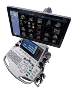
Most commonly associated with pregnancy, this exam uses sounds waves to create images based on patterns returned to a hand-held device. Additional uses include diagnosing infection and cancer and performing biopsy procedures. Unlike X-rays and MRIs, it doesn’t use any radiation
Vascular Ultrasound - This technology enables the physician to see the vascular system and its function, including detection of blood clots.
11 Convenient Houston Locations

506 Medical Center Blvd. Ste. 100
Conroe, TX 77304
P (281) 569-2130
F (281) 763-2609
Services Offered
- Diagnostic CT

22698 Professional Dr. Ste. 150
Kingwood, TX 77339
P (832) 995-5226
F (832) 995-5228
Services Offered
- Diagnostic CT
- MRI

22710 Professional Dr. Ste. 104
Kingwood, TX 77339
P (281) 312-8598
F (281) 312-8599
Services Offered
- PET/CT

22730 Professional Dr.
Kingwood, TX 77339
P (281) 312-8545
F (281) 312-8568
Services Offered
- Diagnostic CT
- Ultrasound
- Biopsy
- Echo

1250 Cypress Station Dr. Ste. B
Houston, TX 77090
P (281) 516-6550
F (281) 516-6551
Services Offered
- Flow Cytometry Lab
- Histology
- PET/CT
- Nuclear Medicine
- UroNav

18488 I-45 South
Shenandoah, TX 77384
P (281) 943-2440
F (281) 943-2404
Services Offered
- Ultrasound
- Echo

129 Vision Park Blvd. Ste. 206
Shenandoah, TX 77384
P (281) 315-8130
F (281) 315-8132
Services Offered
- X-Ray

9319 Pinecroft Dr. Ste. 130
The Woodlands, TX 77380
P (281) 315-8120
F (281) 719-0951
Services Offered

9323 Pinecroft Dr. Ste. 110
The Woodlands, TX 77380
P (281) 943-2440
F (281) 943-2404
Services Offered
- PET/CT
- Diagnostic CT

13700 Medical Complex Dr.
Tomball, TX 77375
P (281) 315-8160
F (281) 315-8161
Services Offered
- Diagnostic CT
- Ultrasound